Copy And Mirror Is Ideal For Single Centrals
I have a love/hate relationship with the Copy and Mirror function in the software. For years it was such a promising tool, but I could never get good initial proposals. Today with the 5.2 Software, the Copy and Mirror function is vastly improved, and I find myself routinely using it when matching single central incisors.
This patient presented with a chief complaint of his poorly matching crown on tooth #9. He has a history of trauma to the tooth as a child and this is his third restoration. This crown has been in service for 10 years, but now the patient would like to replace it for his daughters upcoming wedding (figure 1). As you can see in this retracted view (figure 2) the crown has some thickness issues which is causing the stump to show through. He also has staining at the buccal margin and the shape of the restoration is not perfectly symmetrical with tooth #8.
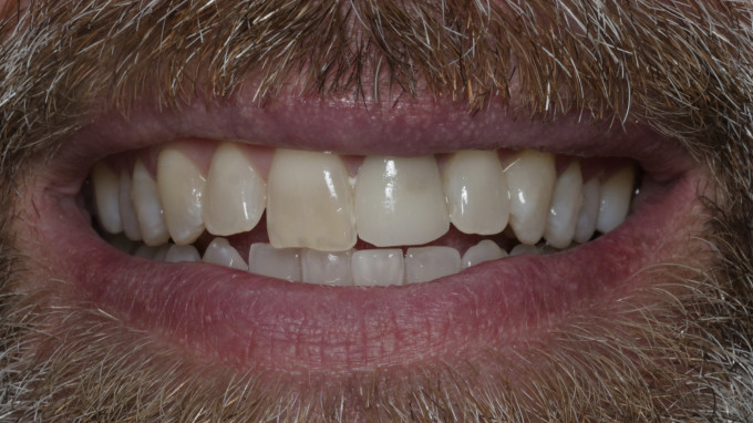
Figure 1: Pre-op condition
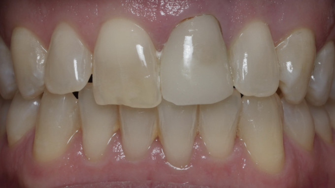
Figure 2: Retracted view
I explained the esthetic challenges with treating a single central versus the entire smile, mainly the inability to change the shade and overall contours of the remaining teeth. He was fairly insistent that he only wanted to replace tooth #9 so that it would match the length and shape of the existing central #8.
After removing the existing ceramic crown, we designated the restoration as a Copy and Mirror and would restore this with Ivoclar e.max (figure 3). You can see how much of the tooth structure was reduced previously, so I was limited in my material choices (figure 4). With this much ceramic thickness, it would be difficult to control the value and match the translucency of his existing tooth. After drawing my margin, I then have the Copy Line step to circumscribe the contralateral tooth, in this case #8 (figure 5).
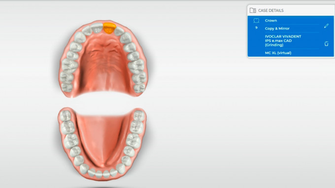
Figure 3: Case Details
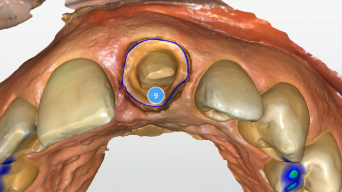
Figure 4: Previous tooth preparation
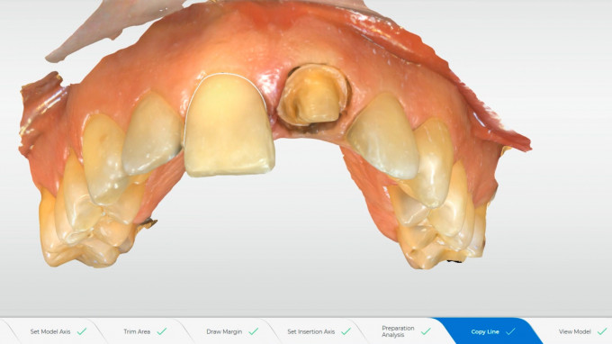
Figure 5: Copy line
The initial proposal was spot on (figure 6). I only had to adjust the mesial and distal contacts using the smooth tool and the restoration was ready to manufacture. At this point I like to show the patients the final proposal and explain what they might expect when it’s delivered. In this case we discussed the gingival embrasure and the potential for a black triangle due to the triangular shape of his centrals. Since I used a 2 cord technique to retract the gingiva, due to the depth of his existing preparations, I was comfortable with the likelihood of some gingival rebound to close the space.
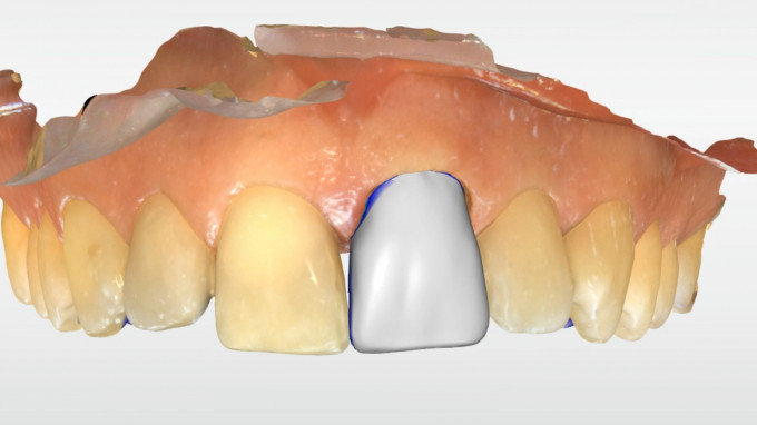
Figure 6: Initial proposal
This case was manufactured using my 4-motor wet/dry MCXL. With the 4 motor milling machines, you have the option of extra find grinding, which produces a finer and truer restoration, with less overmilling and greater anatomical detail. The software will default to fine grinding (figure 7), and once the proper tools are inserted, the extra fine option will be available (figure 8).
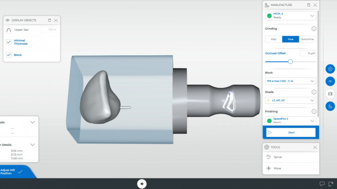
Figure 7: Fine Grinding default
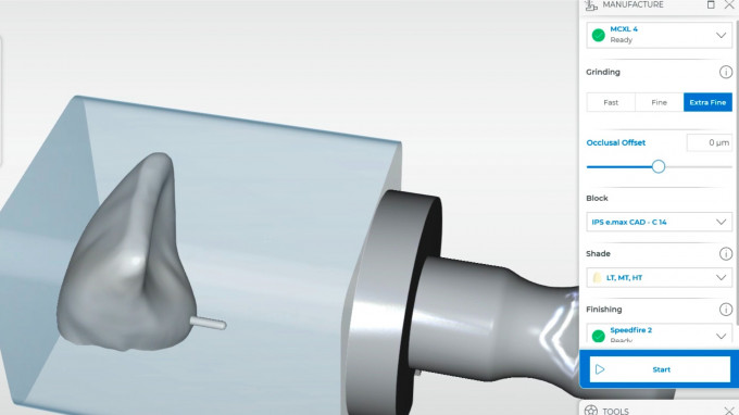
Figure 8: Extra Fine Grinding option
Upon starting the manufacturing job, you may notice the time to be almost twice as much for extra fine (figure 9). However, after the touch process is complete, this time often shrinks down to a more realistic amount. In this case we started with 19:29 and after the touch process we were down to about 13:49 with extra fine grinding.
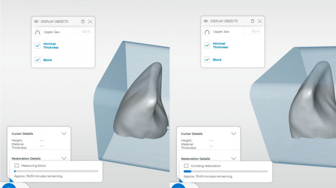
Figure 9: Grinding times
Our shade selection prior to starting was A2-MT. The patient decided to inform me as we were placing the block in the milling machine that he also wants to whiten and wants to make sure his new crown will whiten along with his teeth! So, we decided to increase the value and jump to an A1-MT and allow him to bleach his teeth to match the color.
Here are the 2 week follow up photos (figure 10). You can see the slightly higher value on #9 and the black triangle still present (figure 11). Typically, I wait at least 6-8 weeks for full tissue maturation so I will continue to follow him and post the final photos in a few months once his bleaching has settled in and the tissue rebounds to close the space (figure 12).
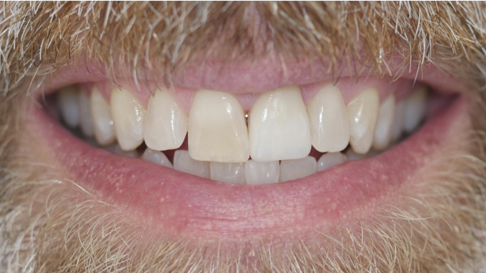
Figure 10: Two week follow-up
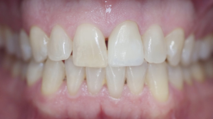
Figure 11: Slight interproximal space
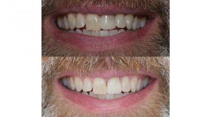
Figure 12: Pre-op vs post-op
Thanks Meena... I almost skipped this with the title, as I have a "love/hate" relationship with that as well, and that's how you started out. I've noticed a difference in the versions over the years, and sometimes it's great, and other's a waste of time. I will always try it if not using a biocopy from a waxup (and that is done in inLab where Copy and MIrror seems a bit better and predicatable), and while it can be effected with some tweeks in model stage, basically it seems to be very good, or a waste of time. GLAD to hear improved in 5.2 which I have not tried in that version yet...Great post
Mark

