Full Mouth Reconstruction Primescan
I am posting this for a couple reasons:
1. This is the first full mouth reconstruction I have done with Primescan. It was amazing and yes MUCH easier than with Omnicam for comprehensive treatment
2. We have has many questions as to what we are doing in our 4B course we have now available. THIS IS ESSENTIALLY IT! How do you execute larger more comprehensive cases with a digital workflow. If you are interested in this, I encourage you to attend. I will be answering any questions you may have on this thread... but nothing replaces a workshop on advanced topics like this.
So some brief background. This patient had two rounds of ortho and orthagnathic surgery. She had previous veneers that were destroyed over the years and with ortho (removing brackets, etc...) and she wanted a new, yes BRIGHT smile, and needed her occlusion restored.
I executed this in two visits....yesterday and today. Exhausting days for sure, but was able to complete it because of proper planning and knowing how to execute it. I just completed it, so the pictures are immediate post op.
I will post the pictures below and we can discuss it however much you all would like:
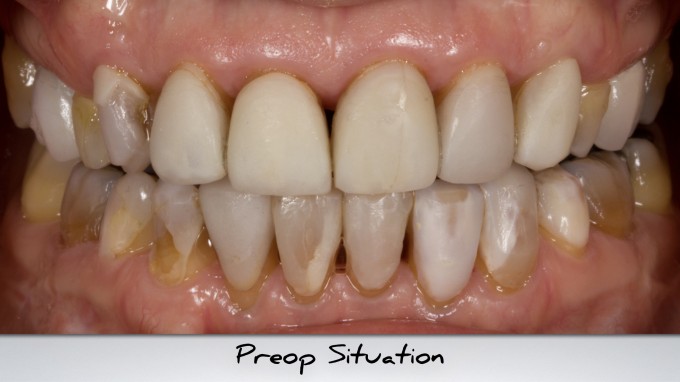
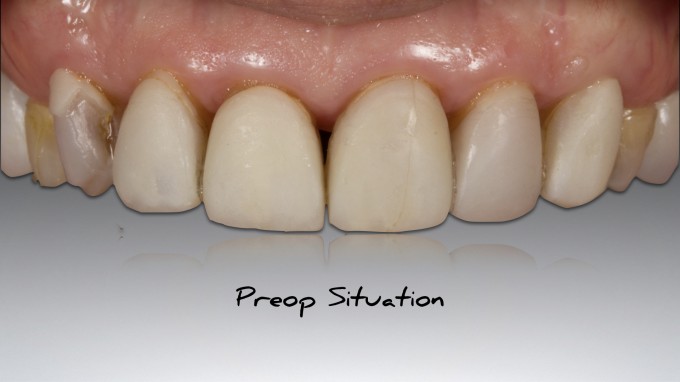
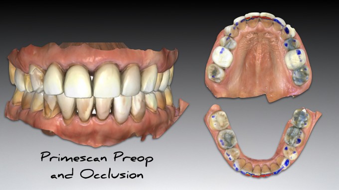
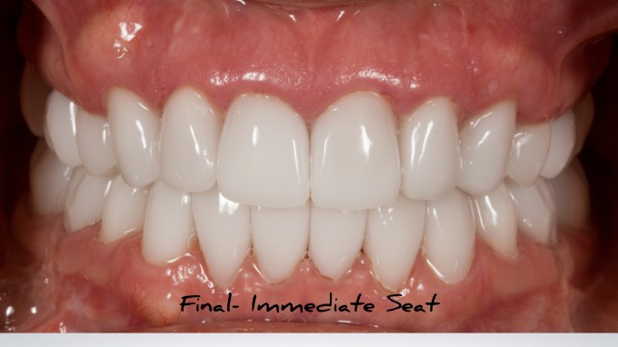
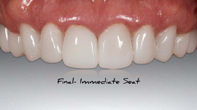
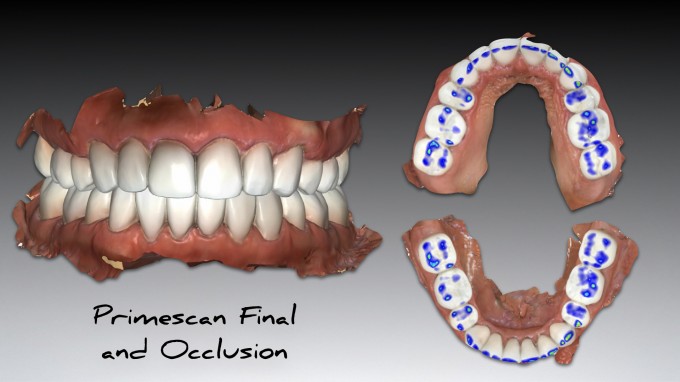
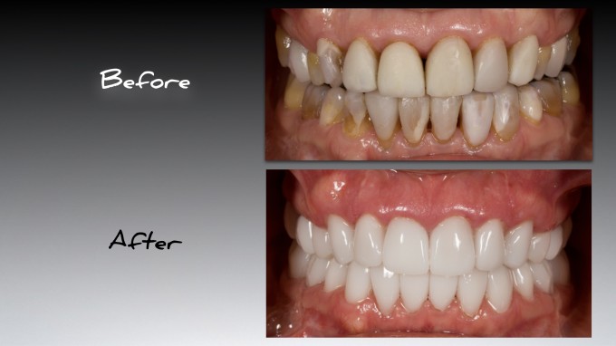
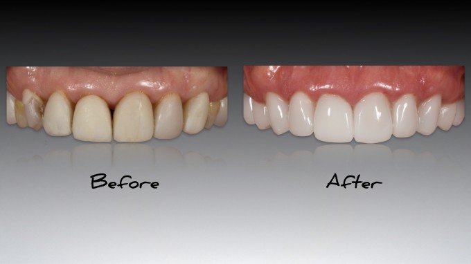
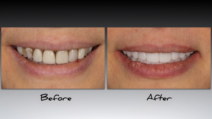
Beautiful work. Obviously all the usual questions pertaining to diagnostics and workflow, but immediate questions for me are how much easier was it to obtain a good full arch scan than over the Omni, and two were there any inaccuracies that you found at try in?
Full arch scan is immensely easier with Primescan... biggest advantage of the scanner. I found very few inaccuracies. In fact there were minimal adjustments needed.
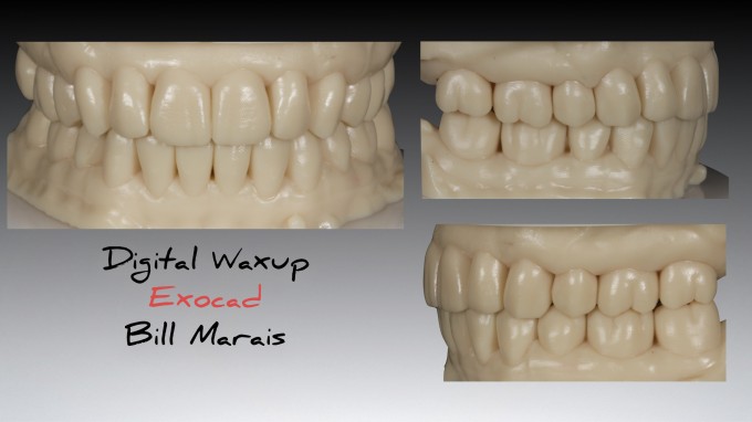
This comes mainly from planning and an excellent, accurate waxup by Bill Marais.
I used the a variation of the “Scottsdale Protocol” we developed for Level 4B and i restores in the following order:
Upper anteriors
Upper posteriors
Lower anteriors
Lower posteriors
Scottsdale Protocol in action. Great work Mike. Textbook execution as taught in the workshop.
Great work as usual. Looking forward to 4b later this month.
Will you be making modifications for the course later this month?
This is starting to look almost too easy, Mike. Great work as always, though! Although it's hard to tell for sure, it appears that Bill did the entire mouth virtual waxup, not just #5-12 and 21-28. Can you walk us through how you stitched your segmental preps to the full-arch waxups without common hard tissue data?
Mike, I do not have Primescan yet, have handled it but not totally familiar with the inner workings. My question is the virtual articulator. Does it operate the same and if so are the measurements from the cone beam golden? I would suppose then getting the final delivery would be without effort to fit the bite.
Peter
Brilliant question Ross... you are going to like this ;)
2 waxups...
first one waxed from 5–12 and 21-28 for segmental Biocopy of anterior...
Then, Bill went into the same file and waxed up molars... can use that one for posteriors after anteriors are done...
I ended up doing Biogeneric on the lowers posterior anyway... but could have done Biocopy. Just wanted to be sure on occlusion and trusted the buccal bite more.
On 3/5/2019 at 6:10 pm, Mike Skramstad said... Brilliant question Ross... you are going to like this ;) 2 waxups... first one waxed from 5–12 and 21-28 for segmental Biocopy of anterior... Then, Bill went into the same file and waxed up molars... can use that one for posteriors after anteriors are done... I ended up doing Biogeneric on the lowers posterior anyway... but could have done Biocopy. Just wanted to be sure on occlusion and trusted the buccal bite more.
Cheater.
Perfect execution of the Scottsdale Protocol. What was the thinking behind using printed models vs the biocopy merge technique?
Merge technique would have taken too much time since it was done all in two visits...better for indirect technique.
Great to see the Primescan and Scottsdale Method in action on a case. WHY was this case done so quickly and would more time have allowed more characterization/anatomy/grinding, or was the patient coming from elsewhere and wanted it done over the shortest period of time? I realize you use the waxup to bring most of the contours into the mouth, but these lack your traditional sharp line angles from the past. They appear more "feminine" and rounded. Not a complaint, just noting the difference, and that I think they look great for this smile.
Hi Ernie-
Good questions.
1. She flew in from California to get the case done... so yes, was schedule to get done in 2 days. She will be in tomorrow morning for final check and then flying home.
2. I had plenty of time and could have added more anatomy if needed (i also would not have gone quite so white), but this patient was extremely particular in what she wanted. We spent two hours in a consult making a list. She wanted them feminine, rounded, not a lot of detail, and glossy ;).
I finished yesterday 2 hours early and today 3 hours early. Time was not a huge issue here. Have to give them what they want... they are paying for it!
Nice case Mike, there's a shocker right?. Look forward to seeing some workflow of this in Charlotte end of the month.
If you had wanted to add the contours, line angles, additional anatomy as Ernie is referencing would you have printed the prep models for use with that or done it live on the patient, or perhaps a PVS impression ( GASP!) for working models. Maybe the 5.0 software has the " virtual red pencil" you taught me for use in visualizing the grind zones?
Amazing case Mike!
Did you find yourself missing the trackball during a long design process such as this? I am still trying to get used to the track pad and often find myself missing the left and right click buttons.
So if we do not have a Bill Marus in our pocket how can we get a wax up like this done? We did a full mouth case a while back that felt a lot like beating my head against a wall while trying to get a digital wax up. What does a lab need to have in place to make this work? I need to come to this course again. So much has changed!!
On 3/6/2019 at 8:43 am, Brett Titensor said...Amazing case Mike!
Did you find yourself missing the trackball during a long design process such as this? I am still trying to get used to the track pad and often find myself missing the left and right click buttons.
I have a wireless mouse that I use for large cases like this. Use the trackpad for day to day stuff
On 3/6/2019 at 9:07 am, Chris Haag said...So if we do not have a Bill Marus in our pocket how can we get a wax up like this done? We did a full mouth case a while back that felt a lot like beating my head against a wall while trying to get a digital wax up. What does a lab need to have in place to make this work? I need to come to this course again. So much has changed!!
Just all the pictures (clinical) and the .stl files of full arch upper and lower is needed. I send them to Bill with instructions and we communicate from there.
I understand, I just recemented a CA veneer that popped off on #6 yesterday. BL-something and opaque cement. I swear those teeth arrive before she does, but that is EXACTLY what she wants and glad a CA dentist gave it to her! I live in the A2 to A4 world, including plenty of C and D shades as well in my practice. Sounds like you tackled the tough stuff with her: having a consult to know the result you wanted BEFORE you start. I do this as well and find not many do.
I have so many questions...
1) When using the scottsdale protocol, we are supposed to scan the wax up models un-preped areas in the main folder to have the data mix a bit before scanning the entire wax up model in the biocopy folder (and pray there is enough info for it to stitch) and wait for the green check mark in the corner. So now with the primescan being so accurate and that it auto cuts the old 'bad' data out, should we still do that little bit of scanning on those untouched molars to allow that bit of mixing of data before scanning in the biocopy folder?
Im more in the boat where I would still be working with stone models and wax on stone models when planning the case. So I would think there is some error in the stone to mouth (more so than printed models to the mouth anyways).
2) Mike, when you say you finished early and dismissed the patient 2 hours early the first day, did you finish essentially the entire upper arch that day? if so, do you worry at all about the occlusion not being harmonious to the lowers (even for 1 night in terms patient chipping the emax restorations over that 1 night)?
3) how many mills and ovens did you have to utilize to be able to make something like that happen in 1 day? even if it was just an upper arch (and I use 'even' and "just" loosely here).
4) are we talking about finishing early in an 8 hour day here or a 10 hour day?
Ernie,
I'm offended by your comment.
We used an A2 block in my office just last week![]()
Mike, Awesome work on a difficult case.
You made it sound easy, but it wasn't
Hi Benson... See your answers below:
On 3/6/2019 at 5:38 pm, Benson Wong said...
I have so many questions...
1) When using the scottsdale protocol, we are supposed to scan the wax up models un-preped areas in the main folder to have the data mix a bit before scanning the entire wax up model in the biocopy folder (and pray there is enough info for it to stitch) and wait for the green check mark in the corner. So now with the primescan being so accurate and that it auto cuts the old 'bad' data out, should we still do that little bit of scanning on those untouched molars to allow that bit of mixing of data before scanning in the biocopy folder?
I still do things about the same way. I had two sets of waxups done and printed. The first waxup had #5-12 and #21-28 waxed up. I used that to do the anterior teeth. That left 4 molars as stitching abutments to correlate the model and the intraoral preps. I prepped 5-12 first and scanned an entire upper arch. I then scanned the model and hoped that the waxup would stitch to the preps. It did not at first so I scanned the molars from the model on top of the molars from the intraoral scan AND scanned the molars from the mouth on top of the molars on the biocopy model... and they stitched.
I did the same thing on day 2 with the lower anterior teeth. This was a bit easier actually.
The second waxup had all the molars waxed up in occlusion. I used this for the upper arch (biocopy) after the Mx Anterior were done and then the lower posteriors in Biogeneric last.
Im more in the boat where I would still be working with stone models and wax on stone models when planning the case. So I would think there is some error in the stone to mouth (more so than printed models to the mouth anyways).
Either one is inaccurate if you transfer the waxup to the mouth in acrylic. Both are quite accurate if you scan the actual model in the Biocopy folder. The Printed models generally will have less detail than the hand waxed ones... but we deal with that due to c0nvenience
2) Mike, when you say you finished early and dismissed the patient 2 hours early the first day, did you finish essentially the entire upper arch that day? if so, do you worry at all about the occlusion not being harmonious to the lowers (even for 1 night in terms patient chipping the emax restorations over that 1 night)?
After we finished the upper arch on day one, I built up the lower molars (off the second waxup model...remember there were 2) with acrylic transferred to the waxup. This stabilized her posterior occlusion over-night. Great question.
3) how many mills and ovens did you have to utilize to be able to make something like that happen in 1 day? even if it was just an upper arch (and I use 'even' and "just" loosely here).
I had 3 mills and 2 ovens running all day.
4) are we talking about finishing early in an 8 hour day here or a 10 hour day?
Day one was 8:00am-3:30am straight through
Day two was 7:30am-2pm straight through
Total time of case: 14 hours
On 3/5/2019 at 5:25 pm, Jeffrey Gregson said... Great work as usual. Looking forward to 4b later this month. Will you be making modifications for the course later this month?
Yes... the entire first day is new... new lab exercises and new lecture material from me. Will be fun . You will be the first course I present my newest material to.
Sweet! Looking forward to seeing what you have in store and having a drink with you guys.
I know you guys go above and beyond with all of this and the work you put into everything. I would like to say thank you for doing so much to pave the way for the rest of us.
See you in a few weeks.
Great Job! Looks amazing!
As a new Cerec user I have a few questions that I haven't seen so far. How did you build the patient's occlusion? Did you build their new occlusion using the Primescan and the wax-ups into Centric Relation? Did you have CR match MIP? (how can you tell I just came from the Spear Occlusion workshop!) Is it hard to get a green check for CR bite records using the primescan?
